Visualizzazione post con etichetta Hematopathology. Mostra tutti i post

martedì 6 gennaio 2015

AN UPDATE IN IMMUNOHISTOCHEMISTRY (PART I and II)

This 2-part special issue features 14 review articles with an attempt to cover IHC automation, standardization of diagnostic IHC, and the role of IHC in diagnosing tumors in major organs and tumors of unknown primary. This series begins with an article emphasizing standardization of diagnostic IHC in the preanalytic, analytic, and postanalytic phases, with a specific focus on (1) newly proposed guidelines on antibody validation from the College of American Pathologists Pathology and Laboratory Quality Center, (2) testing/optimizing a new antibody and troubleshooting, (3) interpreting and reporting IHC assay results, (4) continuing quality improvement programs, and (5) developing and implementing the concept of best practices in IHC.

from Fan Lin (2014) Evolving Practices of Diagnostic Immunohistochemistry. Archives of Pathology & Laboratory Medicine: December 2014, Vol. 138, No. 12, pp. 1561-1563.


Standardization of Diagnostic Immunohistochemistry: Literature Review and Geisinger Experience

Overview of Automated Immunohistochemistry

Immunohistochemistry in Undifferentiated Neoplasm/Tumor of Uncertain Origin

Utility of Immunohistochemistry in the Diagnosis of Pleuropulmonary and Mediastinal Cancers: A Review and Update

Application of Immunohistochemistry in Breast Pathology: A Review and Update

The Application of Immunohistochemical Biomarkers in Urologic Surgical Pathology

New Immunohistochemistry for B-Cell Lymphoma and Hodgkin Lymphoma


Application of Immunohistochemistry in Gastrointestinal and Liver Neoplasms: New Markers and Evolving Practice

Utility of Immunohistochemistry in the Pancreatobiliary Tract

The Utility of Immunohistochemistry in the Differential Diagnosis of Gynecologic Disorders

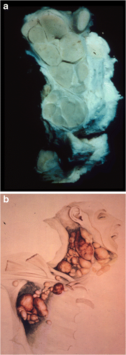
Review and Updates of Immunohistochemistry in Selected Salivary Gland and Head and Neck Tumors

Application of Immunohistochemistry in Thyroid Pathology

Immunohistochemistry in Dermatopathology

An Update on the Application of Newly Described Immunohistochemical Markers in Soft Tissue Pathology

domenica 28 luglio 2013

Thomas Hodgkin: the “man” and “his disease”

Thomas Hodgkin (1798–1865) was one of the leading physicians and scientists of the nineteenth century. His outstanding contributions transcend many fields, medical and non-medical. A renowned diagnostician, he carried out pioneering work in public health and epidemiology, but devoted the greater part of his career to the study of pathology. Among his many interests were anthropology, geology, geography, and ethnography. He helped found the University of London. [...] His most important legacy to medical science was the recognition of the condition called “Hodgkin’s disease,” now known as “Hodgkin lymphoma”. Hodgkin did not fully appreciate the importance of this work and, indeed, it was not recalled as being a particularly noteworthy contribution at the time of his death.

 A un anno dal seminario del prof. Pettinato, posto un recente paper che prende in esame la vita di Thomas Hodgkin e la storia della patologia che da lui ha preso il nome.

Geller SA, Taylor CR. Thomas Hodgkin: the "man" and "his disease": humani nihil a se alienum putabit (nothing human was foreign to him). Virchows Arch. 2013 Jul 26.

a A specimen from the Gordon Museum, Guy’s Hospital of one of the original Hodgkin lymphoma cases described by Hodgkin.


b Water-color drawing by Robert Carswell of case VII . Both images show well the discrete abnormal nodes in contrast to the usual picture of non-Hodgkin lymphoma where nodes are matted together 
(da Geller et al, 2013)

lunedì 17 giugno 2013

Follicular dendritic cells: origin, function, and different disease-associated patterns

Follicular dendritic cells (FDCs) are a specialized type of antigen-presenting dendritic cells that are largely restricted to lymphoid follicles. They form dense three-dimensional meshwork patterns within benign follicles, which maintain the follicular architecture. The FDC function is to bind and retain antigens by linking to complement and immune complexes and then present these antigens to germinal center B cells that start the secondary immune response. FDCs aid in the rescue of bound B cells from apoptosis, and induce the differentiation of B cells into long-term memory B cell clones or plasma cells. We will discuss the different patterns of the FDC meshwork observed in different types of reactive and neoplastic disorders, which may be due to underlying different roles that FDCs may play in these disorders and whether changes in the architecture of the FDC meshwork can be useful in routine diagnostic practice or have a prognostic value.
Rezk SA, Nathwani BN, Zhao X, Weiss LM. Follicular dendritic cells: origin,function, and different disease-associated patterns. Hum Pathol. 2013Jun;44(6):937-50.


FDC patterns in reactive and neoplastic disorders highlighted by CD21 immunostaining, from Rezk et al, 2013.

domenica 9 settembre 2012

Obituary: Karl Lennert

Pubblico un estratto dalla  lettera del prof. Pileri sulla scomparsa di Karl Lennert (la lettera integrale è raggiungibile qui)
Stefano tu sai chi era Tommaso?". Risposi: "Karl, intendi San Tommaso?". Ed Egli disse: "Sì. Non credo, se non vedo!". Ogni giorno, discutemmo i casi rivisti, facendo entrambi un salto all'indietro nel tempo, ritornando al 1980, quando giovane "guest" a Kiel iniziai a confrontarmi con lui, studiando la linfoadenite di Kikuchi, che scoprimmo interessare anche la popolazione Europea. Come allora, i Suoi occhi di un azzurro profondo, che brillavano di luce propria, coglievano i minimi dettagli che, genialmente, rielaborava in principi fisiopatologici. Quei principi che a noi, Suoi allievi, spettava di validare con le tecnologie più avanzate e sofisticate del momento.
Potrei definire Karl un "visionario" nel senso più alto e nobile della parola, perché capace di immense intuizioni, un visionario tuttavia guidato da un rigore metodologico unico, che non si beava mai della propria genialità, ma cercava sempre la prova provata. Ciò che dovrebbe essere ogni vero "Scientist".

domenica 15 aprile 2012

Please, don't shoot the hematocytopathologist!

« Quando un uomo con la pistola incontra un uomo col fucile, quello con la pistola è un uomo morto! »

La sentenza che Ramón Rojo rivolge a Joe lo straniero nel capolavoro di Sergio Leone, è applicabile anche all' ematopatologia? Più precisamente, è sempre vero che quando un patologo con il campione istologico di linfonodo incontra il patologo con l' ago sottile, quest' ultimo è destinato ad arrendersi? 
Il recente editoriale del prof. Zeppa apparso su Cytopathology, rientra in questa annosa polemica aggiungendo un punto di vista interessante.  La varietà delle lesioni accessibili  grazie al prelievo citologico è così vasta che non è impossibile imbattersi, nella routine citologica, in lesioni linfoproliferative extra-nodali. Se il patologo non viene addestrato su questa (non rarissima) evenienza, potrebbe perdere un'occasione diagnostica importante. Si legge nell' editoriale: [lymphoid] cells do not always respect the borders suggested by professional fields and can show the most variable clinical presentations and organ involvement compared with all other forms of human pathology. [...] Hence, they [the cytopathologists] are required to have specific knowledge and professional skills in haematopathology even when they are not directly involved in it. Infine è da ricordare che, anche quando la patologia è localizzata nella più classica sede linfonodale, l' agoaspirato può essere considerato, in particolari situazioni cliniche sfavorevoli al prelievo istologico, come first choice, tenendo conto anche delle enormi potenzialità di tecniche ancillari come la citofluorimetria.

Bibliografia:

Zeppa P. Haematocytopathology: why? Cytopathology. 2012 Apr;23(2):73-5.


martedì 20 dicembre 2011

Lymphoma Classification: from tradition to translation.

Posto il pdf relativo alla lecture del prof. Pileri, che gentilmente ci ha concesso di condividerlo sul nostro blog (LINK).

Il Prof. Karl Lennert e la Kiel Classification (1974) © Pileri 2011.


mercoledì 21 settembre 2011

Pitfalls in Diagnostic Hematopathology

The complexity involved in the histological interpretation of lymph nodes and other lymphoid tissue specimens suspected of harbouring lymphoma is underappreciated [...]  there remains a need to maintain skills in smaller district hospitals, to ensure lymphoma recognition in unexpected circumstances, to permit clinically useful interim diagnoses when needed urgently and to sustain training in haematopathology among junior pathologists. In this review a range of potential pitfalls in lymphoid tissue pathology is outlined, arising at all stages from specimen preparation to reporting. 
(Dall'abstract di Wilkins BS. Pitfalls in lymphoma pathology: avoiding errors in diagnosis of lymphoid tissues)

Bibliografia

Wilkins BS. Pitfalls in lymphoma pathology: avoiding errors in diagnosis of lymphoid tissues. J Clin Pathol. 2011 Jun;64(6):466-76.

Wilkins BS. Pitfalls in bone marrow pathology: avoiding errors in bone marrow trephine biopsy diagnosis. J Clin Pathol. 2011 May;64(5):380-6

Hasserjian RP. Reactive versus neoplastic bone marrow: problems and pitfalls. Arch Pathol Lab Med. 2008 Apr;132(4):587-94