lunedì 26 agosto 2013

Lepidic, a Canadian Neologism

In the new classification of adenocarcinomas, the term lepidic is defined as tumor cells proliferating along the surface of intact alveolar walls without stromal or vascular invasion.
[...] This word is a neologism, a new word invented in Canada in the early 1900s [by] John George Adami, MD. [...] After studying in Manchester, Cambridge, Breslau, and Paris, Dr Adami arrived at McGill University in Montreal, Canada, in 1892. Adami was a prolific writer, and he first used the term lepidic in an address to the Toronto Pathological Society on January 4, 1902. In his lecture titled ‘‘Original Communication on the Classification of Tumors,’’ he proposed 2 new terms that would be used to classify all neoplasms. The term lepidic (from greek meaning a rind, skin, or membrane) was applied to tumors that appeared to be derived from surface-lining cells. The term hylic (from greek meaning crude undifferentiated material) was applied to tumors that appeared to be derived from connective tissues. [...]
[...] The terms lepidic and hylic underwent a subtle change in meaning. In Herbert Spencer’s Pathology of the Lung,6 published in 1962, he stated that tumors may grow into the surrounding alveoli ‘‘either filling them with a solid mass of malignant cells (a hilic [sic] growth) or lining their walls (a lepidic growth).’’ This is a minor alteration in definition; however, this is where the modern definition of tumor growth along intact alveolar septa arose. [...]
In the early 1990s the erroneous belief that the term lepidic came from the etymologic origin ‘‘butterfly’’ became prevalent. [...] Soon the term was stated to mean, ‘‘like a butterfly resting on a branch,’’ ‘‘resembling butterflies sitting on a fence,’’  ‘‘butterflies alighting on shrubbery,’’ ‘‘vague resemblance in cross-section to a butterfly,’’ and a reference ‘‘to the scales of the butterfly wing.’’  While these colorful and useful metaphors evoked both the microscopic morphologic appearance and the possible aerogenous mode of dissemination, they were unrelated to the source of the term. Lepidic did not have an entomologic etymology.

John George Adami (left) first chair of pathology at McGill University (Montreal, Canada) and creator of the term lepidic. Lepidic pattern of adenocarcinoma, characterized by noninvasive surface alveolar growth of tumor cells (right).

Jones KD. Whence Lepidic? Arch Pathol Lab Med. 2013 Aug 12. 

(seen on http://timallenmdjd.blogspot.it/ via Twitter)

domenica 28 luglio 2013

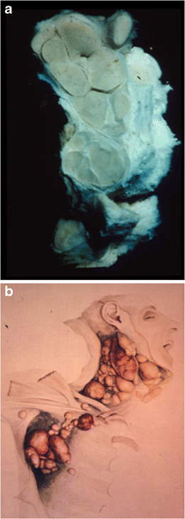
Thomas Hodgkin: the “man” and “his disease”

Thomas Hodgkin (1798–1865) was one of the leading physicians and scientists of the nineteenth century. His outstanding contributions transcend many fields, medical and non-medical. A renowned diagnostician, he carried out pioneering work in public health and epidemiology, but devoted the greater part of his career to the study of pathology. Among his many interests were anthropology, geology, geography, and ethnography. He helped found the University of London. [...] His most important legacy to medical science was the recognition of the condition called “Hodgkin’s disease,” now known as “Hodgkin lymphoma”. Hodgkin did not fully appreciate the importance of this work and, indeed, it was not recalled as being a particularly noteworthy contribution at the time of his death.

 A un anno dal seminario del prof. Pettinato, posto un recente paper che prende in esame la vita di Thomas Hodgkin e la storia della patologia che da lui ha preso il nome.

Geller SA, Taylor CR. Thomas Hodgkin: the "man" and "his disease": humani nihil a se alienum putabit (nothing human was foreign to him). Virchows Arch. 2013 Jul 26.

a A specimen from the Gordon Museum, Guy’s Hospital of one of the original Hodgkin lymphoma cases described by Hodgkin.


b Water-color drawing by Robert Carswell of case VII . Both images show well the discrete abnormal nodes in contrast to the usual picture of non-Hodgkin lymphoma where nodes are matted together 
(da Geller et al, 2013)

domenica 21 luglio 2013

Talking to patients about a diagnosis of malignancy in a FNA clinic setting

Informing patients of a cancer diagnosis is never easy and requires skills not systematically taught in US medical schools and residency programs. The absence of a universally accepted best method for delivering bad news presents an additional challenge. In specialties like pathology, practitioners typically have little patient contact. Training programs, including Cytopathology fellowships, in which patient encounters are routine during fine-needle aspiration (FNA) procedures, offer minimal education on this topic. Formal teaching in this area is lacking, even in medical fields where physicians frequently must present bad news to patients, such as palliative care and oncology.Without adequate training, delivering bad news to patients is made more difficult for physicians in all fields, and the FNA pathologist in particular may be reluctant to participate in patient care beyond obtaining the FNA sample. [...]

Pathologists are integral members of the clinical team, and cytopathologists in particular need to develop communication skills for discussing disease with patients. Deciding whether and how much to tell a patient about the FNA diagnosis is subject to personal judgment and the unique interaction between a patient and pathologist.

Ly, A. (2013), Talking to patients about a diagnosis of malignancy in a fine-needle aspiration clinic setting. Cancer Cytopathology, 121: 339–340. 


lunedì 17 giugno 2013

Follicular dendritic cells: origin, function, and different disease-associated patterns

Follicular dendritic cells (FDCs) are a specialized type of antigen-presenting dendritic cells that are largely restricted to lymphoid follicles. They form dense three-dimensional meshwork patterns within benign follicles, which maintain the follicular architecture. The FDC function is to bind and retain antigens by linking to complement and immune complexes and then present these antigens to germinal center B cells that start the secondary immune response. FDCs aid in the rescue of bound B cells from apoptosis, and induce the differentiation of B cells into long-term memory B cell clones or plasma cells. We will discuss the different patterns of the FDC meshwork observed in different types of reactive and neoplastic disorders, which may be due to underlying different roles that FDCs may play in these disorders and whether changes in the architecture of the FDC meshwork can be useful in routine diagnostic practice or have a prognostic value.
Rezk SA, Nathwani BN, Zhao X, Weiss LM. Follicular dendritic cells: origin,function, and different disease-associated patterns. Hum Pathol. 2013Jun;44(6):937-50.


FDC patterns in reactive and neoplastic disorders highlighted by CD21 immunostaining, from Rezk et al, 2013.

lunedì 3 giugno 2013

A brief chronicle of clinical cytology

Posto un breve excursus sulla storia della citologia clinica apparso recentemente su Diagnostic Cytopathology. Per chi volesse approfondire l' argomento, consiglio l' ottimo libro di Grunze e Spriggs corredato da uno splendido apparato iconografico.

Hampeln, in 1887, published the first case of primary squamous cell carcinoma of the lung and pleura with an illustration of a drawing of cancer cells

(Hampeln P. Über einen Fall von primären Lungen-Pleura-Carinom. St. Petersburg med Wohenschr 1887; 12: 137.)


Bibliografia.


Grunze, H. and A. I. Spriggs (1983). History of clinical cytology: a selection of documents, G-I-T Verlag E. Giebeler.

mercoledì 22 maggio 2013

NIH Director's Wednesday Afternoon Lectures

Dal canale di Youtube del National Institutes of Health, i video dei Seminari del Mercoledì sui più vari aspetti delle scienze biomediche.

The NIH Director's Wednesday Afternoon Lecture Series includes weekly scientific talks by some of the top researchers in the biomedical sciences worldwide.

NIH Youtube Channel (link).

(segnalato dal dr. F. Merolla).


giovedì 9 maggio 2013

Cancer genome landscapes

Over the past decade, comprehensive sequencing efforts have revealed the genomic landscapes of common forms of human cancer. For most cancer types, this landscape consists of a small number of "mountains" (genes altered in a high percentage of tumors) and a much larger number of "hills" (genes altered infrequently). [...] A typical tumor contains two to eight of these "driver gene" mutations; the remaining mutations are passengers that confer no selective growth advantage. Driver genes can be classified into 12 signaling pathways that regulate three core cellular processes: cell fate, cell survival, and genome maintenance. A better understanding of these pathways is one of the most pressing needs in basic cancer research. Even now, however, our knowledge of cancer genomes is sufficient to guide the development of more effective approaches for reducing cancer morbidity and mortality.


(segnalato dal prof. G. Troncone)

Salvador Dalì. Árabes aciddesoxiribonucleics, Paisaje de mariposa. El gran masturbador en paisaje surrealista con ADN.