domenica 22 dicembre 2013

Landmarks in history of cancer

Una serie di articoli che descrive la storia del cancro in rapporto all' evoluzione delle conoscenze mediche.
Una lettura interessante, durante le ferie natalizie.

Bibliografia:


A large scapulohumeral tumor, most likely a sarcoma, was illustrated by Severino in his book, De Recondita, published in 1632. This the first clinical illustration of a tumor.
Hajdu SI. A note from history: landmarks in history of cancer, part 2

sabato 7 dicembre 2013

Ultrasound guided FNA: The new challenges and opportunities for cytopathologists

Numero monografico di Diagnostic Cytopathology dedicato alla categoria degli Interventional Pathologists (vedi post del 24 febbraio 2013). Perchè il patologo non è solo quello che campiona un reperto macroscopico e che legge i preparati al microscopio, ma è sempre più un professionista completo che fa in prima persona il prelievo, visita il paziente e discute la strategie diagnostiche e terapeutiche più appropriate con i colleghi. 

Masood S. Ultrasound guided fine needle aspiration biopsy: The new challenges and opportunities for cytopathologists. Diagn Cytopathol. 2013 Dec;41(12):1017-8.

domenica 17 novembre 2013

Anatomy and Art --- Transforming Vesalius

Un numero monografico della Karger Gazette sul rapporto tra arte e medicina nella storia, in occasione della riedizione critica della Fabrica di Vesalio.

Bibliografia


http://www.vesaliusfabrica.com/, accessed 17/11/2013

Fifth muscle man, displaying the rectus abdominis muscle as it is seen in dogs and caudate apes. Vesalius sometimes altered his woodcuts to show a feature that Galen had described but that does not exist in human anatomy.

domenica 10 novembre 2013

50 ways to diagnose a tumor

Divertente video sulle più improbabili diagnosi differenziali che vengono in mente quando si osserva un caso difficile!

Help me! Help me! These things that I study, are things I'll never see. I've gotta think of every single thing, ' cause everything can look like anything!


sabato 2 novembre 2013

Cineforum 18/11/2013 - Hysteria

La Scuola di Specializzazione in Anatomia Patologica presenta: 

Hysteria

di T. Wexler

con Hugh Dancy, Maggie Gyllenhaal, Felicity Jones, Rupert Everett e Jonathan Pryce.


La proiezione sarà preceduta da un intervento del dott. S. Orlando sull' evoluzione storica del concetto di isteria.

Nota: La proiezione sarà spostata a Lunedì 18 per motivi organizzativi.

Vi aspetto numerosi, ore 15, Auletta Seminari.

domenica 13 ottobre 2013

The Anatomist - by Bill Hayes

La storia dell' Anatomia di Gray, uno dei più famosi libri di anatomia umana in lingua inglese, attraverso i diari e le lettere di Henry Vandyke Carter, il misconosciuto autore del suo splendido corredo iconografico. Bill Hayes, con questo libro, non solo penetra nelle fibre più intime della vita di H.V. Carter ma, attraverso la dissezione in prima persona dei cadaveri, anatomizza anche la propria vita fino a scoprire le più profonde, e toccanti, connessioni fra i vivi e i morti.

What is past - the past - does not, or will it, detach itself and remain where it was (or where it might have been intended to have remained) but it must bring itself forward, and smilingly, or otherwise, present itself as an old friend.

Bill Hayes - The Anatomist: A True Story of Gray's Anatomy.


(libro consigliato dal dr. F. Merolla)

Bibliografia: 

Gray, Henry. Anatomy of the Human Body. Philadelphia: Lea & Febiger, 1918; Bartleby.com, 2000. www.bartleby.com/107/

domenica 29 settembre 2013

International Society of Urological Pathology Consensus Conference on Renal Neoplasia


The International Society of Urological Pathology (ISUP) is the international professional organization dedicated to the subspecialty of urological pathology. [...] In 2011 the Society undertook to review all aspects of the pathology of adult renal malignancy through an international consensus conference [...]. The detailed decisions relating to the consensus conference are presented in 4 reports, included this issue of the journal.
Delahunt B, Egevad L, Montironi R, Srigley JR. International Society of Urological Pathology (ISUP) Consensus Conference on Renal Neoplasia: Rationale and Organization. Am J Surg Pathol. 2013 Oct;37(10):1463-8.

Volume 37(10) pgs. 1463-1633 October 2013

Srigley JR, et al The ISUP Renal Tumor Panel. The International Society of Urological Pathology (ISUP) Vancouver Classification of Renal Neoplasia. Am J Surg Pathol. 2013 Oct;37(10):1469-1489.

Delahunt B,et al, The Members of the ISUP Renal Tumor Panel. The International Society of Urological Pathology (ISUP) Grading System for Renal Cell Carcinoma and Other Prognostic Parameters. Am J Surg Pathol. 2013 Oct;37(10):1490-1504.

Trpkov K, et al; the members of the ISUP Renal Tumor Panel. Handling and Staging of Renal Cell Carcinoma: The International Society of Urological Pathology Consensus (ISUP) Conference Recommendations. Am J Surg Pathol. 2013 Oct;37(10):1505-1517.

Tan PH, et al, The ISUP Renal Tumor Panel. Renal Tumors: Diagnostic and Prognostic Biomarkers. Am J Surg Pathol. 2013 Oct;37(10):1518-1531.

A 69-year-old man, with a history of renal cancer 11 years ago, presented with a 1 cm right lung nodule, which was investigated with fine-needle aspiration under computed tomography guidance. A, Cell block preparation shows tumor cells with pink cytoplasm. B, Higher magnification of tumor cells with pink cytoplasm and nuclei that are vesicular and hyperchromatic. C, Immunohistochemical analysis with RCC marker shows strong cytoplasmic reactivity. D, Pax 2 immunohistochemistry reveals strong nuclear staining, confirming a metastasis to the lung from a primary renal tumor.


Pax 2 and/or Pax 8 were considered to be the most useful markers in the diagnosis of a renal primary.

(from Tan PH, et al, Diagnostic and Prognostic Biomarkers)

P.S. Post n. 100! 

lunedì 23 settembre 2013

Vessels of Stone: Lenin's "Circulatory Disturbance of the Brain"

Many have wondered what might have become of the totalitarian state Lenin founded on merciless terror, had he not died so young. He was 52 and at the height of his power when he had his first stroke. Six months later he had another and then a third stroke three months after that. He died 3 months shy of his 54th birthday with cerebral arteries so calcified that when tapped with tweezers at the time of his autopsy, they sounded like stone. The reason for his premature atherosclerosis has yet to be explained. He had a family history of cardiovascular disease and, therefore, is suspected of having had an inherited lipid disorder. Stress too might have had a role in the progression of his atherosclerosis. However, neither would explain the extent of the calcification of his cerebral arteries identified at post mortem examination. A recently described variant of the NT5E mutation might explain such calcification, as well as Lenin's family history of cardiovascular disease, and his premature cerebrovascular attacks.
Vinters H, Lurie L, Mackowiak PA. Vessels of Stone: Lenin's "Circulatory Disturbance of the Brain". Hum Pathol. 2013 Feb 18.

On the morning of January 23, 1924, the pathologist Alexei Ivanovich Abrikosov was given a task to embalm Lenin's body to keep it intact until the burial. The body is still on permanent display in the Lenin Mausoleum in Moscow. (source Wikipedia)

(to note, Abrikosov has also described the so-called Abrikosov's tumor aka Granular cell tumor)

mercoledì 4 settembre 2013

Looking too closely at thyroid nodules

Endocrinologi e patologi sono certamente co-responsabili dell' incrementata incidenza dei tumori della tiroide. Troppi noduli vengono inviati all' agoaspirato, troppi pazienti subiscono un intervento chirugico e troppe neoplasie a differente comportamento biologico e clinico vengono riunite sotto la stessa terminologia. Un corretto studio clinico può certamente evitare inutili FNA che, talora, vengono utilizzati dai clinici anche in funzione "difensiva". Così come può essere limitata l' indicazione all' intervento chirurgico, applicando agli aspirati i criteri classificativi correnti. Ad esempio, la maggior parte delle citologie "atipiche" (AUS/FLUS) può essere risolta con la semplice ripetizione dell' aspirato (con un risultato il più delle volte di benignità). Diverso il discorso per le lesioni follicolari. Considerata l' incidenza piuttosto bassa del carcinoma follicolare siamo costretti, a causa dei limiti dell' FNA, a mandare consapevolmente molti adenomi follicolari all' intervento chirurgico. Le tecniche di biologia molecolare basate sui profili di espressione genica, aumentano il valore predittivo negativo dell' aspirato "indeterminato", ma non riescono a discriminare (ancora) meglio della morfologia le lesioni effettivamente maligne. Inoltre, sono costose e attualmente non applicabili alla routine clinica. Infine, carcinomi indolenti vengono ancora classificati insieme a quelli a comportamento aggressivo, i cosiddetti "real thyroid carcinomas". Molti carcinomi papillari, come è noto, hanno un comportamento "benigno", come testimoniano i numerosi incidentalomi scoperti nelle sezioni seriate di gozzi nodulari o nelle autopsie di pazienti deceduti per altre cause. Sforzi di correlazione clinica, morfologica e molecolare devono essere fatti per separare i tumori a basso potenziale dai veri carcinomi tiroidei, in modo da ottimizzare efficacemente il managment dei pazienti con patologia nodulare tiroidea.

Bibliografia.


Faquin WC. Can a gene-expression classifier with high negative predictive value solve the indeterminate thyroid fine-needle aspiration dilemma? Cancer Cytopathol. 2013 Mar;121(3):116-9.

Baloch Z, Livolsi VA, Tondon R. Aggressive variants of follicular cell derived thyroid carcinoma; the so called 'Real Thyroid Carcinomas'. J Clin Pathol. 2013 Sep;66(9):733-43.

lunedì 26 agosto 2013

Lepidic, a Canadian Neologism

In the new classification of adenocarcinomas, the term lepidic is defined as tumor cells proliferating along the surface of intact alveolar walls without stromal or vascular invasion.
[...] This word is a neologism, a new word invented in Canada in the early 1900s [by] John George Adami, MD. [...] After studying in Manchester, Cambridge, Breslau, and Paris, Dr Adami arrived at McGill University in Montreal, Canada, in 1892. Adami was a prolific writer, and he first used the term lepidic in an address to the Toronto Pathological Society on January 4, 1902. In his lecture titled ‘‘Original Communication on the Classification of Tumors,’’ he proposed 2 new terms that would be used to classify all neoplasms. The term lepidic (from greek meaning a rind, skin, or membrane) was applied to tumors that appeared to be derived from surface-lining cells. The term hylic (from greek meaning crude undifferentiated material) was applied to tumors that appeared to be derived from connective tissues. [...]
[...] The terms lepidic and hylic underwent a subtle change in meaning. In Herbert Spencer’s Pathology of the Lung,6 published in 1962, he stated that tumors may grow into the surrounding alveoli ‘‘either filling them with a solid mass of malignant cells (a hilic [sic] growth) or lining their walls (a lepidic growth).’’ This is a minor alteration in definition; however, this is where the modern definition of tumor growth along intact alveolar septa arose. [...]
In the early 1990s the erroneous belief that the term lepidic came from the etymologic origin ‘‘butterfly’’ became prevalent. [...] Soon the term was stated to mean, ‘‘like a butterfly resting on a branch,’’ ‘‘resembling butterflies sitting on a fence,’’  ‘‘butterflies alighting on shrubbery,’’ ‘‘vague resemblance in cross-section to a butterfly,’’ and a reference ‘‘to the scales of the butterfly wing.’’  While these colorful and useful metaphors evoked both the microscopic morphologic appearance and the possible aerogenous mode of dissemination, they were unrelated to the source of the term. Lepidic did not have an entomologic etymology.

John George Adami (left) first chair of pathology at McGill University (Montreal, Canada) and creator of the term lepidic. Lepidic pattern of adenocarcinoma, characterized by noninvasive surface alveolar growth of tumor cells (right).

Jones KD. Whence Lepidic? Arch Pathol Lab Med. 2013 Aug 12. 

(seen on http://timallenmdjd.blogspot.it/ via Twitter)

domenica 28 luglio 2013

Thomas Hodgkin: the “man” and “his disease”

Thomas Hodgkin (1798–1865) was one of the leading physicians and scientists of the nineteenth century. His outstanding contributions transcend many fields, medical and non-medical. A renowned diagnostician, he carried out pioneering work in public health and epidemiology, but devoted the greater part of his career to the study of pathology. Among his many interests were anthropology, geology, geography, and ethnography. He helped found the University of London. [...] His most important legacy to medical science was the recognition of the condition called “Hodgkin’s disease,” now known as “Hodgkin lymphoma”. Hodgkin did not fully appreciate the importance of this work and, indeed, it was not recalled as being a particularly noteworthy contribution at the time of his death.

 A un anno dal seminario del prof. Pettinato, posto un recente paper che prende in esame la vita di Thomas Hodgkin e la storia della patologia che da lui ha preso il nome.

Geller SA, Taylor CR. Thomas Hodgkin: the "man" and "his disease": humani nihil a se alienum putabit (nothing human was foreign to him). Virchows Arch. 2013 Jul 26.

a A specimen from the Gordon Museum, Guy’s Hospital of one of the original Hodgkin lymphoma cases described by Hodgkin.


b Water-color drawing by Robert Carswell of case VII . Both images show well the discrete abnormal nodes in contrast to the usual picture of non-Hodgkin lymphoma where nodes are matted together 
(da Geller et al, 2013)

domenica 21 luglio 2013

Talking to patients about a diagnosis of malignancy in a FNA clinic setting

Informing patients of a cancer diagnosis is never easy and requires skills not systematically taught in US medical schools and residency programs. The absence of a universally accepted best method for delivering bad news presents an additional challenge. In specialties like pathology, practitioners typically have little patient contact. Training programs, including Cytopathology fellowships, in which patient encounters are routine during fine-needle aspiration (FNA) procedures, offer minimal education on this topic. Formal teaching in this area is lacking, even in medical fields where physicians frequently must present bad news to patients, such as palliative care and oncology.Without adequate training, delivering bad news to patients is made more difficult for physicians in all fields, and the FNA pathologist in particular may be reluctant to participate in patient care beyond obtaining the FNA sample. [...]

Pathologists are integral members of the clinical team, and cytopathologists in particular need to develop communication skills for discussing disease with patients. Deciding whether and how much to tell a patient about the FNA diagnosis is subject to personal judgment and the unique interaction between a patient and pathologist.

Ly, A. (2013), Talking to patients about a diagnosis of malignancy in a fine-needle aspiration clinic setting. Cancer Cytopathology, 121: 339–340. 


lunedì 17 giugno 2013

Follicular dendritic cells: origin, function, and different disease-associated patterns

Follicular dendritic cells (FDCs) are a specialized type of antigen-presenting dendritic cells that are largely restricted to lymphoid follicles. They form dense three-dimensional meshwork patterns within benign follicles, which maintain the follicular architecture. The FDC function is to bind and retain antigens by linking to complement and immune complexes and then present these antigens to germinal center B cells that start the secondary immune response. FDCs aid in the rescue of bound B cells from apoptosis, and induce the differentiation of B cells into long-term memory B cell clones or plasma cells. We will discuss the different patterns of the FDC meshwork observed in different types of reactive and neoplastic disorders, which may be due to underlying different roles that FDCs may play in these disorders and whether changes in the architecture of the FDC meshwork can be useful in routine diagnostic practice or have a prognostic value.
Rezk SA, Nathwani BN, Zhao X, Weiss LM. Follicular dendritic cells: origin,function, and different disease-associated patterns. Hum Pathol. 2013Jun;44(6):937-50.


FDC patterns in reactive and neoplastic disorders highlighted by CD21 immunostaining, from Rezk et al, 2013.

lunedì 3 giugno 2013

A brief chronicle of clinical cytology

Posto un breve excursus sulla storia della citologia clinica apparso recentemente su Diagnostic Cytopathology. Per chi volesse approfondire l' argomento, consiglio l' ottimo libro di Grunze e Spriggs corredato da uno splendido apparato iconografico.

Hampeln, in 1887, published the first case of primary squamous cell carcinoma of the lung and pleura with an illustration of a drawing of cancer cells

(Hampeln P. Über einen Fall von primären Lungen-Pleura-Carinom. St. Petersburg med Wohenschr 1887; 12: 137.)


Bibliografia.


Grunze, H. and A. I. Spriggs (1983). History of clinical cytology: a selection of documents, G-I-T Verlag E. Giebeler.

mercoledì 22 maggio 2013

NIH Director's Wednesday Afternoon Lectures

Dal canale di Youtube del National Institutes of Health, i video dei Seminari del Mercoledì sui più vari aspetti delle scienze biomediche.

The NIH Director's Wednesday Afternoon Lecture Series includes weekly scientific talks by some of the top researchers in the biomedical sciences worldwide.

NIH Youtube Channel (link).

(segnalato dal dr. F. Merolla).


giovedì 9 maggio 2013

Cancer genome landscapes

Over the past decade, comprehensive sequencing efforts have revealed the genomic landscapes of common forms of human cancer. For most cancer types, this landscape consists of a small number of "mountains" (genes altered in a high percentage of tumors) and a much larger number of "hills" (genes altered infrequently). [...] A typical tumor contains two to eight of these "driver gene" mutations; the remaining mutations are passengers that confer no selective growth advantage. Driver genes can be classified into 12 signaling pathways that regulate three core cellular processes: cell fate, cell survival, and genome maintenance. A better understanding of these pathways is one of the most pressing needs in basic cancer research. Even now, however, our knowledge of cancer genomes is sufficient to guide the development of more effective approaches for reducing cancer morbidity and mortality.


(segnalato dal prof. G. Troncone)

Salvador Dalì. Árabes aciddesoxiribonucleics, Paisaje de mariposa. El gran masturbador en paisaje surrealista con ADN.

sabato 4 maggio 2013

Hypoxic Patterns of Placental Injury

The placenta does not respond in a single way to hypoxia, and various placental hypoxic features should be explained within a clinical context. Because the placenta has a large reserve capacity, hypoxic lesions may not result in poor fetal condition or outcome. On the other hand, very acute, in utero, hypoxic events, followed by prompt delivery, may not be associated with placental pathology, and many poor perinatal outcomes can be explained by an etiology other than hypoxia. Nevertheless, assessment of placental hypoxic lesions is helpful for retrospective explanations of complications in pregnancy and in medicolegal investigation.

Histopathologic features of placental hypoxia da Stanek J. 2013

Stanek J. Hypoxic patterns of placental injury: a review. Arch Pathol Lab Med.2013 May;137(5):706-20.


domenica 7 aprile 2013

Graduated Responsibility for Pathology Residents: No Time for Half Measures

The pathology community is aware of, and has not been idle on, the issue of resident deficiencies. [...] As residency progresses, “[t]he successful trainee develops a very keen sense of dedication, honesty, commitment, intellectual fortitude, integrity, and respect for both patients and colleagues.”Unlike non-pathology residents, who quickly “break the mold formed [in medical school] by being handed a large syllabus containing all of the information that they need to know for an exam and the world of multiple choice questions”… “[i]n contrast, pathology residents may fall into a passive learning style with little or no clinical responsibilities, clinical contact, or accountability…,”leading to “significant deficiencies in confidence, expertise, professionalism, and lifelong learning skills.”
“[a]s the resident progresses over time, the resident may predictate and then give the case to the attending to sign out without sitting with the attending again for sign out. This allows upper level residents to gain additional autonomy”. 
Program directors will need “a clear framework within which to ensure that residents have appropriate opportunities to take responsibility for diagnostic decisions with progressively less oversight.” [...] Program directors will be especially challenged. “[I]t takes a particular combination of high standards, creative thinking, and hard work to manage a training program”; and “time and workload management are critical.” Importantly, “competency-based learning must allow time for remediation for those trainees having inadequate performance…” and “structuring individualized programs for residents in need of remediation falls squarely on the already often overworked residency program director.”


venerdì 29 marzo 2013

Second Opinions: Pathologists' Preventive Medicine

We pathologists have long understood the significance of misdiagnoses and the value of second opinions. Second opinions of pathology diagnoses are routinely used intradepartmentally not only for immediate patient diagnostic accuracy, but also as a tool for peer review, quality assurance, and quality improvement. These may occur as consensus conferences or mandatory second opinions of initial diagnoses of cancers. These measures to assure accuracy of diagnosis are well established in many pathology departments, both private and academic. And directed peer review—selecting specific disease types or particular sites of origin—provides added benefit by targeting latent factors that contribute to diagnostic error.
Da Allen TC. Second Opinions: Pathologists' Preventive Medicine.

Bibliografia:

Allen TC. Second opinions: pathologists' preventive medicine. Arch Pathol Lab Med. 2013 Mar;137(3):310-1.

Landro L. What if the doctor is wrong?: some cancers, asthma, other conditions can be tricky to diagnose, leading to incorrect treatments. Wall Street Journal. January 12,2012 (link) Accessed March 29, 2013.

lunedì 11 marzo 2013

Colorectal cancer: surgical pathology practice guidelines

Caro PATHfinder,

se può essere utile, voglio passarti un piccolo contributo sul cancro del colon.

Ti allego alcuni articoli di Quirke molto didattici che spiegano qual è il metodo e l'obiettivo del patologo nella diagnosi dei tumori del retto. Due articoli spiegano molto chiaramente tutte le possibili lesioni che vanno diagnosticate; un articolo guida nel report macroscopico e microscopico, un altro spiega bene qual è il significato del margine radiale nelle resezioni del retto e come va valutato.
Penso possa essere molto d'aiuto per tutti.

Ciao

Severo Campione, MD.

domenica 24 febbraio 2013

The Interventional Pathologist

We are observing a gradual and important change in the pathologist's profession because a new character has appeared in our daily practice: we can define this character as the “interventional pathologist.” Every day, we now spend most of our time outside of the pathology department: we help nonpathologist physicians and radiologists to solve difficult clinical cases, we aid oncologists in selecting medical procedures, and we verify the adequacy of gastrointestinal biopsies in the endoscopy room. We also discuss the best procedure for molecular characterization of neoplasms. Broadly speaking, we give an effective, direct, and crucial contribution to the patients' diagnostic and therapeutic workup. Moreover, in our hospitals, pathologists personally perform cytologic biopsies, such as thyroid or lymph node fine-needle aspiration biopsy, and histologic sampling, such as breast core needle biopsy. Therefore, we have to interact with radiologists for correct localization of target lesions. [...] This expanded approach to the practice of pathology requires a unique multidisciplinary knowledge. For example, we believe that the pathologist should have a basic knowledge of imaging, and be able to interpret radiography and ultrasound images. [...] The interventional pathologist becomes a modern and all-encompassing figure in the diagnostic process. Are we ready for this cultural revolution?

Bibliografia.


lunedì 11 febbraio 2013

The development of visual search expertise in pathology residents

As pathology is a visual specialty, it would seem that to optimize training, an understanding of  how visual perception skills develop and change as a function of experience would be beneficial.
Insomma, nel corso della specializzazione,  i giovani patologi passano gradualmente dal navigare in maniera "caotica" attraverso il vetrino fino ad arrivare, verso gli ultimi anni, a fermarsi e zoomare solo sulle aree di interesse diagnostico. E tutto questo è possibile solo con l' esposizione quotidiana a casi numerosi e diversificati, sotto una costante supervisione di seniors e professori unita ad uno studio puntuale e rigoroso. Proprio quello che avviene tutti i giorni della nostra residency...

La "semplificazione" progressiva dei movimenti oculari dal I al IV anno di specializzazione. Da Krupinski et al, Human Pathology 2013.


Bibliografia:

domenica 10 febbraio 2013

Don't miss the gynecopathology free issue!

Il numero free di Histopathology dedicato ai più recenti topics sulla patogenesi e la diagnostica delle neoplasie del tratto genitale femminile.

Histopathology, January 2013, Annual review issue: gynaecological pathology.

martedì 29 gennaio 2013

Surgical Pathology Snapshot #10: a 1.600 years old woman


Woman, aged 30-40 at the time of the death.

What's your diagnosis?


domenica 20 gennaio 2013

Dissection: a rite of passage

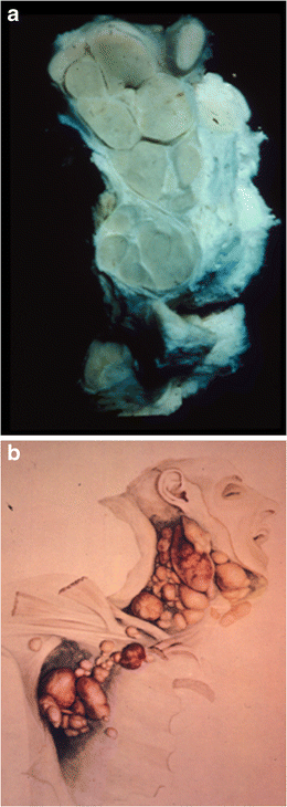
La pratica settoria su cadavere è stata per molto tempo fondamentale negli studi di medicina. Come è accaduto per i riscontri diagnostici, anche la pratica anatomica ha risentito dei tempi attuali, dove  le ricostruzioni digitali hanno via via eliminato la necessità di disporre concretamente di materiale cadaverico. Ma c'era un tempo in cui il corso di anatomia rappresentava un rito di passaggio, che i giovani studenti americani immortalavano all' alba dell' era fotografica. Quei giovani studenti con abiti antiquati, morti anch' essi come i loro soggetti di studio, ci osservano intorno ai cadaveri su cui facevano pratica di anatomia. Era un' epoca ai confini della morale e della legge, dove i cosiddetti resurresionists (i body snatchers di cui abbiamo visto le gesta romanzate nel primo cineforum) rifornivano le scuole di medicina con cadaveri per lo più di condannati, poveri ed afro-americani. La segregazione razziale continuava anche dopo la morte, ed in numerose fotografie si possono osservare futuri medici, prevalentemente bianchi, sezionare cadaveri, prevalentemente neri. Al Museum of London e' in corso  una mostra sul recente ritrovamento,  nel cimitero del Royal London Hospital, di numerosi scheletri risalenti a fine '800 con evidenti segni di pratiche settorie, testimonianza archeologica dell' attività dei resurresionists e della loro intima relazione con le scuole di medicina e chirurgia.  Fra le memorie italiane di questo "rito di passaggio", l' unica che ricordo è quella del poeta Salvatore di Giacomo, un' esperienza terribile che, fortunatamente per noi, lo ha strappato agli studi di medicina. Ecco di seguito la sua testimonianza autobiografica apparsa nell' Occhialetto di Napoli nel 1886:
Nello scorcio d’un malinconico ottobre, una mattina, tra le otto e le nove, mi avviavo  lentamente alla lezione d’anatomia, su per le scale di Sant’Aniello a Caponapoli. Pioviccicava; era una pioggerella diaccia e sottile che penetrava le ossa, una di quelle tristezze lagrimose con cui il cielo diventato grigio principia a piangere l’estate che se ne va. La scala era deserta e muta; [...]. Con le mani in saccoccia, il sigaro inumidito tra le labbra, soffrendo orribilmente a cagione d’uno stivalino troppo stretto, io salivo quel calvario dei miei diciott’ anni e pigliavo per l’ anfiteatro anatomico, col buon volere che avrebbe mosso un condannato a far la via del patibolo. Non so bene se ancora esista  quell' indegna cantina della morte, dove i cadaveri degli Incurabili si sciorinavano in tutta la miseria delle loro carni. [...]. I morti arrivati di fresco dormono in quella stanzetta a man destra, nudi, stesi supini, in una sacrilega confusione di sesso. [...]. Mettete l’ occhio all' inferriata d’un finestrino; ecco là, sulla panca bruttata di sangue, un cadavere giovanile, con le braccia penzoloni, la testa rasa, rovesciata. [...] Se avete coraggio, spingete ancora un usciolino in fondo: questa che segue è la bottega delle ossa; gli studenti vi possono comperare un paio di stinchi, un lucido cranio, una mandibola, una rotella, l’ umano bambù d 'una spina dorsale..
   Non so come, non so perché io scesi, in quella mattina, assalito da un indefinibile presentimento, la scala del deposito. Entrai, di mala voglia, nella sala in cui il professore anatomizzava, e mi posi in mezzo ai compagni miei, che guardavano, con attenzione grande  il cadavere d'un vecchio. Poco prima, seppi, uno di loro s'era svenuto. Era un novizio, un abruzzese, gentile e non forte. Ma come il professore aveva conciato quel vecchio morto!... [...]. Al meglio della lezione, uscii dalla sala. Non ne potevo più; mi si rivoltava lo stomaco. Senza guardarmi attorno, senza salutare nessuno, infilai il corridoio e feci per ascendere, in fretta e furia, la scaletta. In cima un bidello si preparava a discendere, con in capo una tinozza di membra umane. I gradini della scaletta, su per i quali erano passate centinaia di scarpe goccianti, parevano insaponati. Il bidello scivolò, la tinozza - Dio mio! - la tinozza rovesciata sparse per la scala il suo contenuto, e, in un attimo, tre o quattro teste mazze, inseguite da gambe sanguinanti, saltarono per la scala fino a' miei piedi! Di sopra il bidello urlava e sacramentava, raggomitolato in un angolo, aferrandosi una gamba lussata...
Quell’ inserviente, dalla faccia butterata e cinico, dall’ aria insolente, dalla voce sempre rauca, com' egli era sempre oscenamente avvinazzato, si chiamava Ferdinando. Per la faccia sua, cincischiata a quel modo, i compagni lo chiamavano, napoletanamente, Setaccio. lo devo la mia salvazione a Setaccio, perché da quel giorno la cantina dei cadaveri non mi vide più e nemmanco l’ Università, dove compivo il terzo anno di medicina.
Dissection at the Yale School of Medicine around 1910. From the book Dissection.


Bibliografia:

domenica 6 gennaio 2013

Discovered on (but not restricted to) GIST

L' Anoctamin-1 (ANO1) è una proteina canale del cloro calcio-attivata inizialmente descritta nei GIST, nota anche con il nome di Discovered on Gist 1 (DOG1). L' anticorpo commerciale anti-DOG1 è quindi  entrato rapidamente a far parte della famiglia dei markers per i GIST ed è utile soprattutto nei rari GIST negativi per cKIT. Studi successivi hanno però mostrato come l' espressione di questa proteina non fosse limitata ai soli GIST. Infatti DOG1 rappresenta un marker di differenziazione acinare sia nelle ghiandole salivari che nel pancreas ed è stato osservato inoltre anche nei condroblastomi. Ma nulla e più preoccupante, dal punto di vista diagnostico differenziale, dell' espressione di DOG1 in un subset di leiomiomi, sarcomi sinoviali e leiomiosarcomi, in questi ultimi anche (raramente) in associazione con il cKIT. Questo ci ricorda la necessità di adoperare sempre con cautela le nostre armi immunoistochimiche, da utilizzare preferibilmente in associazione e inscrivendole nel più ampio quadro clinico e morfologico.

Bibliografia:


Chênevert J, et al.  DOG1: a novel marker of salivary acinar and intercalated duct differentiation. Mod Pathol. 2012 Jul;25(7):919-29.


Sah SP, McCluggage WG. DOG1 immunoreactivity in uterine leiomyosarcomas. J Clin Pathol. 2013 Jan;66(1):40-3.


giovedì 3 gennaio 2013

Patologie presepiali

Restando ancora per qualche giorno in tema natalizio, vorrei proporvi le immagini di due pastori del diciottesimo secolo. L' arte presepiale, soprattutto nel '700, era caratterizzata da un (iper)realismo grottesco e barocco con cui venivano rappresentate scene e personaggi della Napoli dei vicoli e delle campagne circostanti. Molto spesso gli artisti indugiavano nella rappresentazione dei difetti, e quindi non poteva mancare anche quella delle condizioni patologiche, soprattutto di patologie così diffuse sul nostro territorio come quelle della tiroide. L' ampio ma diffuso ingrossamento della tiroide della figura giovane è più probabilmente dovuto ad una patologia benigna tipo gozzo, mentre la tumefazione monolaterale della vecchia, che sembra anche aver perso peso, potrebbe essere un segno di patologia maligna. E voi, cosa ne pensate?

Le figure fanno parte della collezione della Harvey Cushing/John Hay Whitney Medical Library della Yale University di New Haven, Connecticut e furono donate da Mr. Anthony Van Wagenen figlio della dottoressa Gertrude Van Wagenen, una pioniera nel campo della endocrinologia della riproduzione.


Fonte: