martedì 6 gennaio 2015

AN UPDATE IN IMMUNOHISTOCHEMISTRY (PART I and II)

This 2-part special issue features 14 review articles with an attempt to cover IHC automation, standardization of diagnostic IHC, and the role of IHC in diagnosing tumors in major organs and tumors of unknown primary. This series begins with an article emphasizing standardization of diagnostic IHC in the preanalytic, analytic, and postanalytic phases, with a specific focus on (1) newly proposed guidelines on antibody validation from the College of American Pathologists Pathology and Laboratory Quality Center, (2) testing/optimizing a new antibody and troubleshooting, (3) interpreting and reporting IHC assay results, (4) continuing quality improvement programs, and (5) developing and implementing the concept of best practices in IHC.

from Fan Lin (2014) Evolving Practices of Diagnostic Immunohistochemistry. Archives of Pathology & Laboratory Medicine: December 2014, Vol. 138, No. 12, pp. 1561-1563.


Standardization of Diagnostic Immunohistochemistry: Literature Review and Geisinger Experience

Overview of Automated Immunohistochemistry

Immunohistochemistry in Undifferentiated Neoplasm/Tumor of Uncertain Origin

Utility of Immunohistochemistry in the Diagnosis of Pleuropulmonary and Mediastinal Cancers: A Review and Update

Application of Immunohistochemistry in Breast Pathology: A Review and Update

The Application of Immunohistochemical Biomarkers in Urologic Surgical Pathology

New Immunohistochemistry for B-Cell Lymphoma and Hodgkin Lymphoma


Application of Immunohistochemistry in Gastrointestinal and Liver Neoplasms: New Markers and Evolving Practice

Utility of Immunohistochemistry in the Pancreatobiliary Tract

The Utility of Immunohistochemistry in the Differential Diagnosis of Gynecologic Disorders

Review and Updates of Immunohistochemistry in Selected Salivary Gland and Head and Neck Tumors

Application of Immunohistochemistry in Thyroid Pathology

Immunohistochemistry in Dermatopathology

An Update on the Application of Newly Described Immunohistochemical Markers in Soft Tissue Pathology

Journal Club #11: Human cancer immunotherapy with antibodies to the PD-1 and PD-L1 pathway

The programmed death 1 (PD-1) receptor and its ligands programmed death ligand 1 (PD-L1) and PD-L2, members of the CD28 and B7 families, play critical roles in T cell coinhibition and exhaustion. Overexpression of PD-L1 and PD-1 on tumor cells and tumor-infiltrating lymphocytes, respectively, correlates with poor disease outcome in some human cancers. Monoclonal antibodies (mAbs) blockading the PD-1/PD-L1 pathway have been developed for cancer immunotherapy via enhancing T cell functions. Clinical trials with mAbs to PD-1 and PDL1 have shown impressive response rates in patients, particularly for melanoma, non-small-cell lung cancer (NSCLC), renal cell carcinoma (RCC), and bladder cancer. Further studies are needed to dissect the mechanisms of variable response rate, to identify biomarkers for clinical response, to develop small-molecule inhibitors, and to combine these treatments with other therapies.

Ohaegbulam KC, Assal A, Lazar-Molnar E, Yao Y, Zang X. Human cancer immunotherapy with antibodies to the PD-1 and PD-L1 pathway. Trends Mol Med. 2015 Jan;21(1):24-33.


Human cancer immunotherapy with anti-programmed death 1 (PD-1) receptor and anti-programmed death ligand 1 (PD-L1)/L2 antibodies (from Ohaegbulam et al).    
          

lunedì 1 dicembre 2014

domenica 16 novembre 2014

Journal Club #9: Focal myositis, an intramuscular mass-like reactive process.

Focal myositis is an uncommon inflammatory pseudotumor of skeletal muscle that can be confused with a variety of neoplastic and inflammatory diseases. It is often misunderstood because it presents as a tumor-like mass, but histologically resembles a skeletal muscle myopathy or dystrophy. [...] Histologically, these were solitary intramuscular processes composed of variable myopathic and focal neurogenic changes, fibrosis, and inflammation, occasionally accompanied by prominent eosinophils.[...] Clinical diagnostic considerations ranged from benign entities such as rhabdomyoma, intramuscular lipoma, fibromatosis, myositis ossificans, proliferative myositis, inflammatory myofibroblastic tumor, and inflammatory myopathy to malignant entities such as rhabdomyosarcoma, leiomyosarcoma, liposarcoma, and lymphoma. Available follow-up revealed spontaneous regression.[...] Careful attention to reproducible clinicopathologic features can aid diagnosis and spare patients from excessive surgery or adverse therapy.


Bibliography.

Layfield LJ, Crim J, Gupta D. Fine-needle aspiration findings in nodular myositis: a case report. Diagn Cytopathol. 2000 Nov;23(5):343-7

martedì 29 luglio 2014

Lost in Pathology

Lo so. Il blog langue da un bel po (l' ultimo post risale al 22 aprile!). Qualcuno l' ha sicuramente dato per morto, altri avranno detto: ecco, la solita iniziativa che comincia bene e poi....
Francesco Merolla mi ha dato l' occasione per un post estivo, segnalandomi un interessante numero della rivista Nature sulle Key Advances in Medicine, in cui si riassumono gli hot topics della ricerca medica, corredati da una selezione di paper pubblicati sulle riviste del gruppo Nature nel 2013. Una interessante ma agile lettura estiva! 
Dopo il (meritato) riposo, i post sul blog riprenderanno, con una auspicabile partecipazione di specializzandi, dottorandi e docenti. Da fine settembre riprenderanno inoltre gli incontri del Journal Club, che hanno riscosso un buon successo in termini di partecipazione. E poi i Seminari, il Cineforum e numerose altre iniziative per vivacizzare la formazione degli specializzandi e non.
Perché, nel nostro (meraviglioso) mestiere, non si finisce mai di imparare!

Buone vacanze,

Pathfinder.


Key Advances in Medicine, January 2014, Nature Reviews.

martedì 22 aprile 2014

Journal Club #3: H. pylori detected by PCR-based technique.

PCR-based techniques may substitute the hystochemical and immunohistochemical stainings in the detection of H. pylori in gastric biopsy. Moreover, these new assays can identify mutations that predict antibiotic resistance, avoiding the time-consuming coltures.

Bibliography.

Schmitt BH, Regner M, Mangold KA, Thomson RB Jr, Kaul KL. PCR detection of clarithromycin-susceptible and -resistant Helicobacter pylori from formalin-fixed, paraffin-embedded gastric biopsies. Mod Pathol. 2013 Sep;26(9):1222-7.

Weiss J, Tsang TK, Meng X, Zhang H, Kilner E, Wang E, Watkin W. Detection of Helicobacter pylori gastritis by PCR: correlation with inflammation scores and immunohistochemical and CLOtest findings. Am J Clin Pathol. 2008 Jan;129(1):89-96.

Yantiss RK, Lamps LW. To stain or not to stain...That remains the question. Am J Clin Pathol. 2012 Mar;137(3):343-5. 
Comment on
Hartman DJ, Owens SR. Are routine ancillary stains required to diagnose Helicobacter infection in gastric biopsy specimens? An institutional quality assurance review. Am J Clin Pathol. 2012 Feb;137(2):255-60.


martedì 25 marzo 2014

Journal Club #2 - Tumor budding as prognostic marker in colorectal cancer

Tumor budding (defined as single neoplastic cells or clusters up to five cells at the invasive front of CRC) may represent a new prognostic marker in colorectal cancer. In particular, it could be useful to indentify those pT3N0M0 "high risk" patients who may benefit of additional therapeutic regimen.
Interestingly, tumor budding can be assessed also in pre-operative biopsy and in cancers other than CRCs. 

Overview of the histomorphological and molecular features of the tumour centre, invasive front and tumour buds in CRC. Reproduced from Zlobec and Lugli, 2010.


Bibliography.

Wang LM, Kevans D, Mulcahy H, O'Sullivan J, Fennelly D, Hyland J, O'Donoghue D, Sheahan K. Tumor budding is a strong and reproducible prognostic marker in T3N0 colorectal cancer. Am J Surg Pathol. 2009 Jan;33(1):134-41.


Rogers AC, Gibbons D, Hanly AM, Hyland JM, O'Connell PR, Winter DC, Sheahan K. Prognostic significance of tumor budding in rectal cancer biopsies before neoadjuvant therapy. Mod Pathol. 2014 Jan;27(1):156-62.

Giger OT, Comtesse SC, Lugli A, Zlobec I, Kurrer MO. Intra-tumoral budding in preoperative biopsy specimens predicts lymph node and distant metastasis in patients with colorectal cancer. Mod Pathol. 2012 Jul;25(7):1048-53.

Sarioglu S, Acara C, Akman FC, Dag N, Ecevit C, Ikiz AO, Cetinayak OH, Ada E; for Dokuz Eylül Head and Neck Tumour Group (DEHNTG). Tumor budding as a prognostic marker in laryngeal carcinoma. Pathol Res Pract. 2010 Feb 15;206(2):88-92.

Liang F, Cao W, Wang Y, Li L, Zhang G, Wang Z. The prognostic value of tumor budding in invasive breast cancer. Pathol Res Pract. 2013 May;209(5):269-75.


giovedì 13 febbraio 2014

FNA is the ideal tool to study and analyze tumor heterogeneity at metastatic sites

In routine practice, suspected metastases in patients with cancer are only occasionally biopsied, primarily because of the cost and invasiveness of the procedure. However, biopsies of metastatic lesions can be valuable, not only in confirming the presence of metastatic disease, but also in revealing unsuspected benign disease or secondary malignancies. In addition, such biopsies also allow the assessment of biomarkers that might differ from those on primary tumor cells, and can thereby facilitate selection of the optimal treatment. Because of the increasing recognition of clonal and phenotypic heterogeneity of tumors, we anticipate that in the near future, biopsying of metastatic lesions will constitute a standard-of-care practice, allowing assessment of molecular differences between the primary tumor and metastatic lesions. In our opinion, fine-needle aspiration is currently the best method for making repeated biopsies to monitor the tumor: it is minimally invasive, safe, and cost effective and can be coupled with modern ancillary techniques. Here we provide an up-to-date review of the clinical implications of tumor heterogeneity in metastatic disease and the ancillary molecular techniques used in cytology; we also discuss the role of modern cytology in contemporary diagnosis and management of metastatic cancer.
Beca F, Schmitt F. Growing indication for FNA to study and analyze tumor heterogeneity at metastatic sites. Cancer Cytopathol. 2014 Jan 29.

mercoledì 1 gennaio 2014

Getting Out From Behind the Paraffin Curtain

A must-to-read Editorial on pathologist's career with a focus on the political efforts needed  to achieve a good work quality and the consideration deserved among clinician colleagues and hospital administrators. To get out from behind the paraffin curtain.

Many pathologists, especially those who maintain contracts with hospitals and other providers, possess keen political skills. They have learned how to keep clinicians happy with the accuracy and alacrity of laboratory services, administrators satisfied with efficiency and financial performance, and technical personnel content with working conditions. [...] As pathologists' careers develop, they are often called on to be leaders. Some are uncomfortable in leadership positions, their careers to date having been founded mostly on individual achievement. The reluctant leader should not be dissuaded by lack of leadership experience. Often the best leaders are those who did not seek out the position but developed their own style as they adapted to the demands of the position. [...] Other challenges include learning to delegate (and follow up on delegated tasks), providing encouragement and inspiration to team members (some of whom may be quite hard-boiled and cynical), and occasionally standing up for the team when it is under assault by powerful factions, including clinicians and other clients. This last challenge takes no small measure of courage and equanimity. [...] The pathologist/politician/leader is commonly confronted by ethical issues. We typically serve multiple masters, including patients, administrators, clinicians, and regulatory agencies. We need to be sensitive to their conflicting agendas. Medical ethics is a complex field [...]  Nevertheless, building a reputation for incorruptibility does foster trust, and trust is fertile ground for opportunity.
Edward O. Uthman (2014) Getting Out From Behind the Paraffin Curtain. Archives of Pathology & Laboratory Medicine: January 2014, Vol. 138, No. 1, pp. 12-13.

Happy New Year 2014.

domenica 22 dicembre 2013

Landmarks in history of cancer

Una serie di articoli che descrive la storia del cancro in rapporto all' evoluzione delle conoscenze mediche.
Una lettura interessante, durante le ferie natalizie.

Bibliografia:


A large scapulohumeral tumor, most likely a sarcoma, was illustrated by Severino in his book, De Recondita, published in 1632. This the first clinical illustration of a tumor.
Hajdu SI. A note from history: landmarks in history of cancer, part 2

sabato 7 dicembre 2013

Ultrasound guided FNA: The new challenges and opportunities for cytopathologists

Numero monografico di Diagnostic Cytopathology dedicato alla categoria degli Interventional Pathologists (vedi post del 24 febbraio 2013). Perchè il patologo non è solo quello che campiona un reperto macroscopico e che legge i preparati al microscopio, ma è sempre più un professionista completo che fa in prima persona il prelievo, visita il paziente e discute la strategie diagnostiche e terapeutiche più appropriate con i colleghi. 

Masood S. Ultrasound guided fine needle aspiration biopsy: The new challenges and opportunities for cytopathologists. Diagn Cytopathol. 2013 Dec;41(12):1017-8.

domenica 17 novembre 2013

Anatomy and Art --- Transforming Vesalius

Un numero monografico della Karger Gazette sul rapporto tra arte e medicina nella storia, in occasione della riedizione critica della Fabrica di Vesalio.

Bibliografia


http://www.vesaliusfabrica.com/, accessed 17/11/2013

Fifth muscle man, displaying the rectus abdominis muscle as it is seen in dogs and caudate apes. Vesalius sometimes altered his woodcuts to show a feature that Galen had described but that does not exist in human anatomy.

domenica 10 novembre 2013

50 ways to diagnose a tumor

Divertente video sulle più improbabili diagnosi differenziali che vengono in mente quando si osserva un caso difficile!

Help me! Help me! These things that I study, are things I'll never see. I've gotta think of every single thing, ' cause everything can look like anything!


sabato 2 novembre 2013

Cineforum 18/11/2013 - Hysteria

La Scuola di Specializzazione in Anatomia Patologica presenta: 

Hysteria

di T. Wexler

con Hugh Dancy, Maggie Gyllenhaal, Felicity Jones, Rupert Everett e Jonathan Pryce.


La proiezione sarà preceduta da un intervento del dott. S. Orlando sull' evoluzione storica del concetto di isteria.

Nota: La proiezione sarà spostata a Lunedì 18 per motivi organizzativi.

Vi aspetto numerosi, ore 15, Auletta Seminari.

domenica 13 ottobre 2013

The Anatomist - by Bill Hayes

La storia dell' Anatomia di Gray, uno dei più famosi libri di anatomia umana in lingua inglese, attraverso i diari e le lettere di Henry Vandyke Carter, il misconosciuto autore del suo splendido corredo iconografico. Bill Hayes, con questo libro, non solo penetra nelle fibre più intime della vita di H.V. Carter ma, attraverso la dissezione in prima persona dei cadaveri, anatomizza anche la propria vita fino a scoprire le più profonde, e toccanti, connessioni fra i vivi e i morti.

What is past - the past - does not, or will it, detach itself and remain where it was (or where it might have been intended to have remained) but it must bring itself forward, and smilingly, or otherwise, present itself as an old friend.

Bill Hayes - The Anatomist: A True Story of Gray's Anatomy.


(libro consigliato dal dr. F. Merolla)

Bibliografia: 

Gray, Henry. Anatomy of the Human Body. Philadelphia: Lea & Febiger, 1918; Bartleby.com, 2000. www.bartleby.com/107/. 

domenica 29 settembre 2013

International Society of Urological Pathology Consensus Conference on Renal Neoplasia


The International Society of Urological Pathology (ISUP) is the international professional organization dedicated to the subspecialty of urological pathology. [...] In 2011 the Society undertook to review all aspects of the pathology of adult renal malignancy through an international consensus conference [...]. The detailed decisions relating to the consensus conference are presented in 4 reports, included this issue of the journal.
Delahunt B, Egevad L, Montironi R, Srigley JR. International Society of Urological Pathology (ISUP) Consensus Conference on Renal Neoplasia: Rationale and Organization. Am J Surg Pathol. 2013 Oct;37(10):1463-8.

Volume 37(10) pgs. 1463-1633 October 2013

Srigley JR, et al The ISUP Renal Tumor Panel. The International Society of Urological Pathology (ISUP) Vancouver Classification of Renal Neoplasia. Am J Surg Pathol. 2013 Oct;37(10):1469-1489.

Delahunt B,et al, The Members of the ISUP Renal Tumor Panel. The International Society of Urological Pathology (ISUP) Grading System for Renal Cell Carcinoma and Other Prognostic Parameters. Am J Surg Pathol. 2013 Oct;37(10):1490-1504.

Trpkov K, et al; the members of the ISUP Renal Tumor Panel. Handling and Staging of Renal Cell Carcinoma: The International Society of Urological Pathology Consensus (ISUP) Conference Recommendations. Am J Surg Pathol. 2013 Oct;37(10):1505-1517.

Tan PH, et al, The ISUP Renal Tumor Panel. Renal Tumors: Diagnostic and Prognostic Biomarkers. Am J Surg Pathol. 2013 Oct;37(10):1518-1531.

A 69-year-old man, with a history of renal cancer 11 years ago, presented with a 1 cm right lung nodule, which was investigated with fine-needle aspiration under computed tomography guidance. A, Cell block preparation shows tumor cells with pink cytoplasm. B, Higher magnification of tumor cells with pink cytoplasm and nuclei that are vesicular and hyperchromatic. C, Immunohistochemical analysis with RCC marker shows strong cytoplasmic reactivity. D, Pax 2 immunohistochemistry reveals strong nuclear staining, confirming a metastasis to the lung from a primary renal tumor.


Pax 2 and/or Pax 8 were considered to be the most useful markers in the diagnosis of a renal primary.

(from Tan PH, et al, Diagnostic and Prognostic Biomarkers)

P.S. Post n. 100! 

lunedì 23 settembre 2013

Vessels of Stone: Lenin's "Circulatory Disturbance of the Brain"

Many have wondered what might have become of the totalitarian state Lenin founded on merciless terror, had he not died so young. He was 52 and at the height of his power when he had his first stroke. Six months later he had another and then a third stroke three months after that. He died 3 months shy of his 54th birthday with cerebral arteries so calcified that when tapped with tweezers at the time of his autopsy, they sounded like stone. The reason for his premature atherosclerosis has yet to be explained. He had a family history of cardiovascular disease and, therefore, is suspected of having had an inherited lipid disorder. Stress too might have had a role in the progression of his atherosclerosis. However, neither would explain the extent of the calcification of his cerebral arteries identified at post mortem examination. A recently described variant of the NT5E mutation might explain such calcification, as well as Lenin's family history of cardiovascular disease, and his premature cerebrovascular attacks.
Vinters H, Lurie L, Mackowiak PA. Vessels of Stone: Lenin's "Circulatory Disturbance of the Brain". Hum Pathol. 2013 Feb 18.

On the morning of January 23, 1924, the pathologist Alexei Ivanovich Abrikosov was given a task to embalm Lenin's body to keep it intact until the burial. The body is still on permanent display in the Lenin Mausoleum in Moscow. (source Wikipedia)

(to note, Abrikosov has also described the so-called Abrikosov's tumor aka Granular cell tumor)

mercoledì 4 settembre 2013

Looking too closely at thyroid nodules

Endocrinologi e patologi sono certamente co-responsabili dell' incrementata incidenza dei tumori della tiroide. Troppi noduli vengono inviati all' agoaspirato, troppi pazienti subiscono un intervento chirugico e troppe neoplasie a differente comportamento biologico e clinico vengono riunite sotto la stessa terminologia. Un corretto studio clinico può certamente evitare inutili FNA che, talora, vengono utilizzati dai clinici anche in funzione "difensiva". Così come può essere limitata l' indicazione all' intervento chirurgico, applicando agli aspirati i criteri classificativi correnti. Ad esempio, la maggior parte delle citologie "atipiche" (AUS/FLUS) può essere risolta con la semplice ripetizione dell' aspirato (con un risultato il più delle volte di benignità). Diverso il discorso per le lesioni follicolari. Considerata l' incidenza piuttosto bassa del carcinoma follicolare siamo costretti, a causa dei limiti dell' FNA, a mandare consapevolmente molti adenomi follicolari all' intervento chirurgico. Le tecniche di biologia molecolare basate sui profili di espressione genica, aumentano il valore predittivo negativo dell' aspirato "indeterminato", ma non riescono a discriminare (ancora) meglio della morfologia le lesioni effettivamente maligne. Inoltre, sono costose e attualmente non applicabili alla routine clinica. Infine, carcinomi indolenti vengono ancora classificati insieme a quelli a comportamento aggressivo, i cosiddetti "real thyroid carcinomas". Molti carcinomi papillari, come è noto, hanno un comportamento "benigno", come testimoniano i numerosi incidentalomi scoperti nelle sezioni seriate di gozzi nodulari o nelle autopsie di pazienti deceduti per altre cause. Sforzi di correlazione clinica, morfologica e molecolare devono essere fatti per separare i tumori a basso potenziale dai veri carcinomi tiroidei, in modo da ottimizzare efficacemente il managment dei pazienti con patologia nodulare tiroidea.

Bibliografia.


Faquin WC. Can a gene-expression classifier with high negative predictive value solve the indeterminate thyroid fine-needle aspiration dilemma? Cancer Cytopathol. 2013 Mar;121(3):116-9.

Baloch Z, Livolsi VA, Tondon R. Aggressive variants of follicular cell derived thyroid carcinoma; the so called 'Real Thyroid Carcinomas'. J Clin Pathol. 2013 Sep;66(9):733-43.

lunedì 26 agosto 2013

Lepidic, a Canadian Neologism

In the new classification of adenocarcinomas, the term lepidic is defined as tumor cells proliferating along the surface of intact alveolar walls without stromal or vascular invasion.
[...] This word is a neologism, a new word invented in Canada in the early 1900s [by] John George Adami, MD. [...] After studying in Manchester, Cambridge, Breslau, and Paris, Dr Adami arrived at McGill University in Montreal, Canada, in 1892. Adami was a prolific writer, and he first used the term lepidic in an address to the Toronto Pathological Society on January 4, 1902. In his lecture titled ‘‘Original Communication on the Classification of Tumors,’’ he proposed 2 new terms that would be used to classify all neoplasms. The term lepidic (from greek meaning a rind, skin, or membrane) was applied to tumors that appeared to be derived from surface-lining cells. The term hylic (from greek meaning crude undifferentiated material) was applied to tumors that appeared to be derived from connective tissues. [...]
[...] The terms lepidic and hylic underwent a subtle change in meaning. In Herbert Spencer’s Pathology of the Lung,6 published in 1962, he stated that tumors may grow into the surrounding alveoli ‘‘either filling them with a solid mass of malignant cells (a hilic [sic] growth) or lining their walls (a lepidic growth).’’ This is a minor alteration in definition; however, this is where the modern definition of tumor growth along intact alveolar septa arose. [...]
In the early 1990s the erroneous belief that the term lepidic came from the etymologic origin ‘‘butterfly’’ became prevalent. [...] Soon the term was stated to mean, ‘‘like a butterfly resting on a branch,’’ ‘‘resembling butterflies sitting on a fence,’’  ‘‘butterflies alighting on shrubbery,’’ ‘‘vague resemblance in cross-section to a butterfly,’’ and a reference ‘‘to the scales of the butterfly wing.’’  While these colorful and useful metaphors evoked both the microscopic morphologic appearance and the possible aerogenous mode of dissemination, they were unrelated to the source of the term. Lepidic did not have an entomologic etymology.

John George Adami (left) first chair of pathology at McGill University (Montreal, Canada) and creator of the term lepidic. Lepidic pattern of adenocarcinoma, characterized by noninvasive surface alveolar growth of tumor cells (right).

Jones KD. Whence Lepidic? Arch Pathol Lab Med. 2013 Aug 12. 

(seen on http://timallenmdjd.blogspot.it/ via Twitter)

domenica 28 luglio 2013

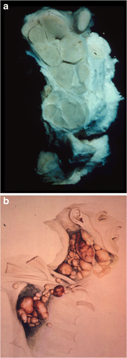
Thomas Hodgkin: the “man” and “his disease”

Thomas Hodgkin (1798–1865) was one of the leading physicians and scientists of the nineteenth century. His outstanding contributions transcend many fields, medical and non-medical. A renowned diagnostician, he carried out pioneering work in public health and epidemiology, but devoted the greater part of his career to the study of pathology. Among his many interests were anthropology, geology, geography, and ethnography. He helped found the University of London. [...] His most important legacy to medical science was the recognition of the condition called “Hodgkin’s disease,” now known as “Hodgkin lymphoma”. Hodgkin did not fully appreciate the importance of this work and, indeed, it was not recalled as being a particularly noteworthy contribution at the time of his death.

 A un anno dal seminario del prof. Pettinato, posto un recente paper che prende in esame la vita di Thomas Hodgkin e la storia della patologia che da lui ha preso il nome.

Geller SA, Taylor CR. Thomas Hodgkin: the "man" and "his disease": humani nihil a se alienum putabit (nothing human was foreign to him). Virchows Arch. 2013 Jul 26.

a A specimen from the Gordon Museum, Guy’s Hospital of one of the original Hodgkin lymphoma cases described by Hodgkin.


b Water-color drawing by Robert Carswell of case VII . Both images show well the discrete abnormal nodes in contrast to the usual picture of non-Hodgkin lymphoma where nodes are matted together 
(da Geller et al, 2013)